Mr Nikolaos Panagiotopoulos PhD FACS FICS FSSO
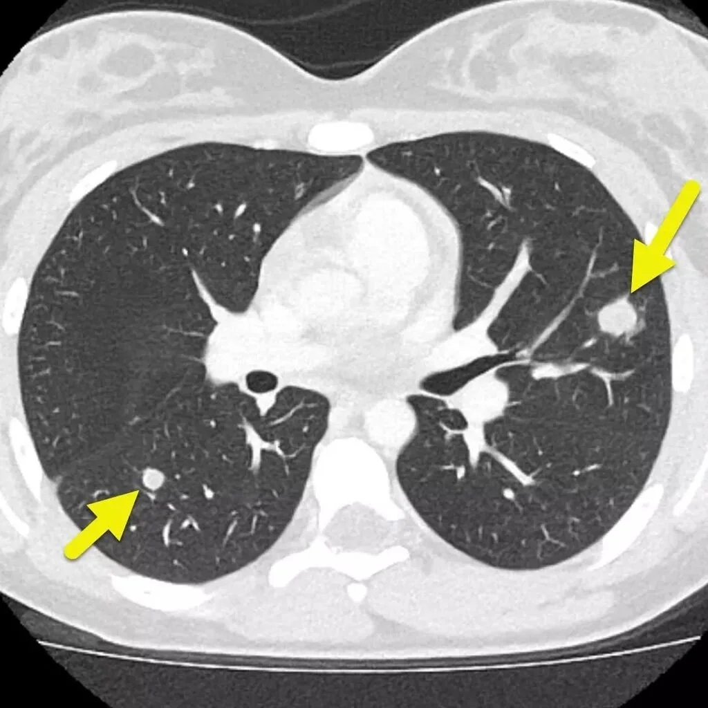
Thoracic and Robotic SurgeonWhat is a Pulmonary Nodule?A pulmonary nodule (also known as a lung nodule) is a small, round or oval spot in the lung, typically measuring less than 3 cm, seen on a CT scan or chest X-ray.Most pulmonary nodules are benign (non-cancerous), but a small proportion may represent early-stage lung cancer, which is why proper evaluation and follow-up are essential.With the increasing use of CT imaging, lung nodules are now frequently detected incidentally during scans performed for other reasons. 1. Solid Pulmonary NodulesAppear as dense, well-defined lesions on CTMost common typeCan be benign (e.g. scar tissue, granuloma) or malignantRisk depends on size, margins, and growth over time
2. Subsolid Nodules• Ground-glass nodules (GGN)Appear hazy and less dense than solid nodulesMay represent inflammation, infection, or early lung adenocarcinoma
• Part-solid nodulesContain both solid and ground-glass componentsHigher risk of malignancy compared to pure ground-glass nodules
Navigating the Management Pathway
From watchful observation to active intervention — each step is guided by evidence and tailored to the individual patient.
-
For small or benign-appearing nodules, regular CT scans monitor for growth or change over time. Scan intervals are determined by nodule size and patient risk profile.
-
When a nodule appears suspicious or demonstrates growth, tissue sampling is required. Options include percutaneous lung biopsy (CT-guided needle), Endobronchial Ultrasound (EBUS) for airway-adjacent nodules, Robotic Navigational Bronchoscopy ION for lesions located deep in the lung parenchyma
-
with relevant specialities including oncologist, radiologist, respiratory physician
-
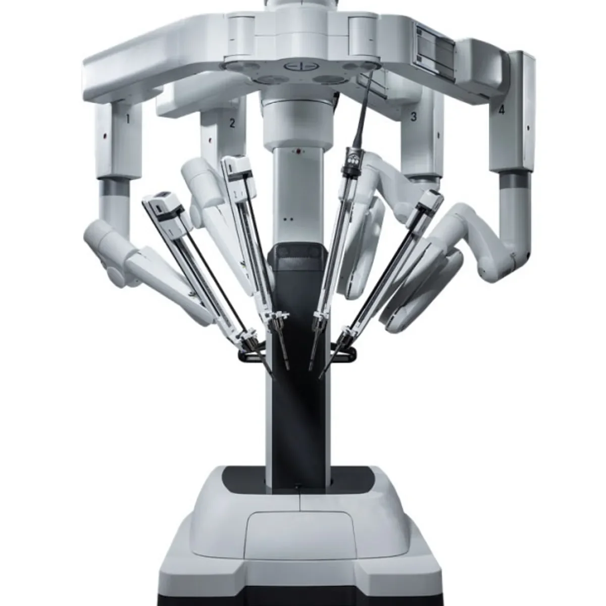
Confirmed malignancy may be managed with surgical resection, stereotactic body radiotherapy (SBRT), or percutaneous ablation.
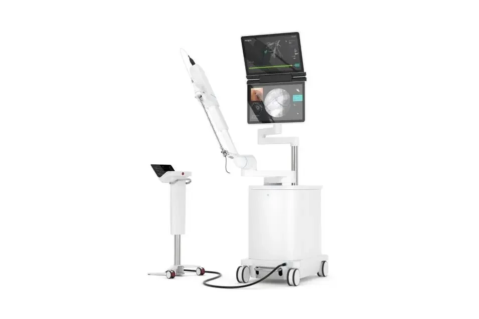
Lung Nodules: Early Diagnosis with Robotic Navigational BronchoscopyLung nodules are increasingly detected on routine CT scans—often incidentally and in patients without symptoms. While most nodules are benign, some may represent early-stage lung cancer, making accurate and timely diagnosis essential.Traditionally, investigating small or deep lung nodules has been challenging. Standard bronchoscopy often cannot reach peripheral lesions, and CT-guided biopsies carry risks such as pneumothorax.Robotic Navigational Bronchoscopy (RNB) is transforming how we approach lung nodule investigation.Using advanced 3D CT mapping and robotic-assisted precision, RNB allows us to:
✔ Reach small and difficult-to-access nodules
✔ Obtain accurate biopsies with high diagnostic yield
✔ Minimise complications compared to traditional techniques
✔ Diagnose lung cancer at an earlier, more treatable stageThis technology represents a major step forward in early lung cancer detection and personalised care.Mr Nikolaos Panagiotopoulos MD PhD FSSO FICS
Consultant Thoracic and Robotic Surgeon
Cleveland Clinic London
33 Grosvenor Place, SW1 7HY London UK
Tel: 0044 (0) 2036337963
Mobile: 0044 (0) 7752 194604
Email: nick@chestsurgeon.co.uk